Cone Beam CT
As Albert Einstein said, “If I can’t picture it, I can’t understand it.”
Sleep and Brain aims to identify a disease process, establish a differential diagnosis, determine an etiology, and render treatment options to ensure optimal health. We gather as much information as possible through a comprehensive exam, data collection, and photography. Radiographic imaging using cone beam computed tomography (CBCT) offers insight into causes and treatment solutions by visualizing anatomy. CBCT provides detailed images of the jaw, teeth, upper airway, nasal cavity, and sinuses. The best clinical outcomes occur when we can see what is happening.
Horizontal Dimension
Children and adults can display horizontal dimension deficiencies, commonly seen in sleep-related disordered breathing due to improper growth of the maxilla and mandible. Maxillary deficiency enables the soft palate to restrict the airway. Similarly, a retrognathic mandible forces the tongue back into the throat and pharyngeal region, constricting the airway. Early interventions can correct and prevent these outcomes and their effects on health.
Vertical Dimension
Vertical dimension is the distance between any point on the maxilla and the mandible when the teeth are in maximum intercuspation. Aberrancies in the vertical dimension are associated with sleep and brain disorders.
Imaging is essential to recognize problems in the temporomandibular joint region, vital in airway management and treating TMD. Active or passive joint degeneration and disk injury result in vertical dimension loss, causing pain and function loss in the joint and head and neck muscles.
This vertical dimensional loss can also impact occlusion and teeth wear. The CBCT images can indicate the need for additional imaging, like an MRI, to understand better the damage to the temporal mandibular disk. A loss of vertical dimension can also negatively impact the airway. When considering a mandibular advancement appliance to treat obstructive sleep apnea, it is crucial to understand disk position and the presence of active or passive condylar degeneration since compressive forces can exacerbate these conditions.
The ramus length dictates the lower facial one-third and changes the vertical dimension. With normal ramus development, the facial thirds are similar in length. With short ramus development, a long lower facial one-third with an increased display of teeth and gum on the maxillary arch develops. A long ramus has the opposite appearance. Instead of a long, narrow face, a more square face grows. Treatment for short ramus may entail double jaw surgery to decrease the lower facial one-third and decrease the teeth amount displayed on the maxillary arch. In contrast, the opposite is valid for a long ramus.
The gonial angle also influences the vertical dimension. An acute gonial angle (flat mandibular plane angle) will mimic the facial appearance of a long ramus. In contrast, an obtuse gonial angle (steep mandibular plane angle) will mimic the facial appearance of a short ramus length. The strength of the masseter muscles influences the gonial angle. The stronger and more developed the masseter muscle is, the more acute the gonial angle will be.
Transverse Dimension
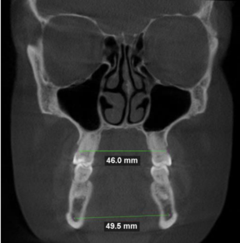
Transverse deficiencies result in sleep disorders, malocclusion, tooth wear, and improper growth. We can measure the maxilla and mandible to understand their growth relationship.
Compared to the mandible, the ideal relationship would be a maxilla plus 5-10 mm measurement. The image also demonstrates the patient's inability to get their tongue to the palate. How the tongue fills this space indicates a narrow arch, possibly precipitated by a tongue-tie or childhood mouth breathing, which affected growth. Recognizing transverse dimension issues early can produce predictable corrections through expansion. TAD expanders, TPD, and EASE can correct transverse deficiency in children and adults, improving their occlusion, sleep, and overall brain health.
Nasal Anatomical Abnormalities
Nasal anatomical abnormalities increase nasal resistance and reduce nasal flow, leading to mouth breathing.
A CBCT detects a deviated septum, bone spur, and enlarged turbinates due to allergies or concha bullosa. Proper nasal breathing is critical to maximizing brain health, correcting mouth breathing, and improving sleep quality. The images help determine if noninvasive treatments like nasal saline rinse, mouth tape, nasal dilators, allergy regimens, or Buteyko breathing may be effective. The photos also help a head and neck or maxillofacial surgeon visualize a surgical plan to correct the anatomy.
Airway Volume
Imaging the airway space is beneficial to understanding the impact of the soft tissue and its effect on airflow. CBCT identifies potential chokepoints, which are the areas where the tissue can close off, impairing breathing.
\
Typically, an average airway volume is 150 mm; less than that would raise the suspicion for sleep-disordered breathing.
Tonsils and Adenoids
A CBCT identifies the presence or absence of enlarged tonsils and adenoids.
Enlarged tonsillar tissue impacts sleep and brain health by narrowing, restricting, or blocking the airway. Identifying airway impingement is especially important with children, as early intervention improves growth and development.
Frequently asked questions
-
Planmeca Viso® G7 utilizes Planmeca Ultra Low Dose™ – a proprietary low-dose 3D imaging protocol that enables CBCT imaging with an even lower radiation dose than standard panoramic imaging. This protocol allows us to acquire 3D images at significantly lower effective doses without statistically reducing image quality. The pioneering and scientifically proven protocol is based on intelligent low-dose 3D imaging algorithms that produces an optimal balance of dose and quality – helping us adhere to the ALADA principle (As Low As Diagnostically Acceptable). SCARA (Selectively Compliant Articulated Robot Arm) technology guarantees anatomically accurate imaging geometry. The SCARA arm can produce any required movement pattern. This technology enables accurate and reliable volume positioning and diameter adjustment, resulting in clear and error-free CBCT images and reducing the dosage exposure. Finally, the Planmeca AINO™ algorithm achieves noise-free CBCT images without losing valuable details. The algorithm improves image quality when using small voxel sizes and allows lowering exposure values by reducing noise.
-
Movement artifacts are among the most significant challenges to CBCT image quality. When a subject moves during imaging, it produces artifacts that affect the quality of the image. Planmeca Viso® G7 utilizes the intelligent Planmeca CALM® movement correction algorithm to cancel the effects caused by this movement. The Planmeca CALM® algorithm is excellent for imaging restless children, individuals with special needs, or the elderly. The Planmeca CALM® algorithm saves time and guards patients against unnecessary exposures by eliminating the need for retakes by canceling the effects of movement. Additionally, Planmeca Viso® G7’s head support provides stability and comfort. The clever support design of Planmeca Viso® G7 does not cover the ears in a 3D face photograph. We can also use it without the chin cup thanks to the Planmeca CALM® movement artifact correction algorithm.
-
Metal restorations and root fillings can cause shadows and streaks in dental CBCT images. Based on extensive scientific research, the Planmeca Viso® G7 utilizes the intelligent Planmeca ARA™ algorithm to remove these artifacts efficiently and reliably.
-
Rhinomanometry detects the Aerodynamic Failure (the traffic jam). The CBCT reveals the Architectural Cause (the car crash). When resistance is high, we use the 3D Scan to identify which of the four specific barriers is restricting flow inside the nose:
Septal Deviation: Is the center cartilage bent or spurred, physically blocking one lane of the airway?
Turbinate Obstruction: We check the structures that heat your air for two distinct issues: Hypertrophy (permanent soft-tissue swelling) or Concha Bullosa (a hidden air pocket inside the bone itself that steals valuable airspace).
Transverse Deficiency Is your upper jaw (maxilla) too narrow? If the "floor" of the nose is constricted, the entire nasal tunnel is structurally cramped. This often requires orthopedic expansion rather than just surgery.
Adenoids & Polyps: We check for growths that standard exams often miss. Nasal Polyps can grow out of the sinuses to fill the airway tunnel, while enlarged Adenoid Tissue can physically plug the "back exit" of the nose (the choanae).


